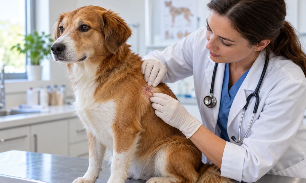
How veterinarians diagnose mast cell tumors in dogs begins with a physical exam and a sample from the suspicious lump. A bump’s size, color, or softness cannot reliably show whether it is cancer. Many mast cell tumors resemble harmless growths, and some swell or shrink temporarily. If you are researching an antihistamine for mast cell tumors in dogs, first ask your veterinarian whether a tumor has been confirmed and what the medication is meant to do. Antihistamines may be part of a veterinary plan, but they do not diagnose or remove a tumor.
Start with the history and physical examination
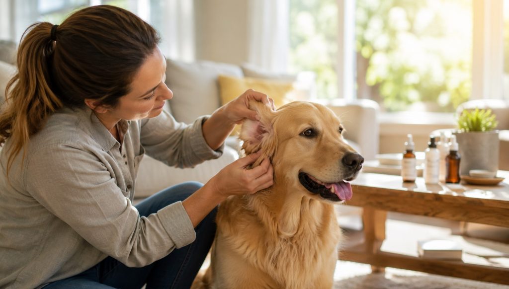
The veterinarian will ask when the lump appeared, how it has changed, and whether your dog licks or scratches it. Mention any swelling, redness, bruising, bleeding, or tenderness. Bring photos taken over time, a list of medications and supplements, and details about appetite, energy, vomiting, or stool changes. Tell the clinic if your dog has several lumps or a previous mast cell tumor.
During the exam, the veterinarian measures and palpates the mass and checks the surrounding skin. They may examine other lumps and feel nearby lymph nodes. These observations help choose the next steps, but touch alone cannot confirm a mast cell tumor. Even experienced clinicians need laboratory evaluation to distinguish it from other skin growths.
Fine needle aspiration is often the first test
A fine needle aspirate uses a small needle to collect cells from the mass. The veterinarian spreads the sample on a slide for microscopic review. Mast cells often have characteristic features, so cytology can identify many tumors without removing the whole lump. The procedure is usually brief, though some dogs may need help staying still.
Sometimes the first sample is too small, contains mostly blood, or does not provide enough detail. The veterinarian may recommend repeating the aspirate, sampling another area, or sending slides to a veterinary pathologist. Ask how confident the result is and whether the next step is monitoring, biopsy, or removal.
How veterinarians diagnose mast cell tumors in dogs can involve more than identifying the cells. A cytology result may confirm a mast cell tumor, but it does not always predict how aggressive it is. Tissue analysis after biopsy or surgery can provide information about grade and other features used to guide prognosis and treatment.
Biopsy and histopathology
A biopsy removes a small piece of tissue for analysis. In other cases, a surgeon removes the whole mass with margins planned for the location and size. A pathologist examines the tissue under a microscope. The report may describe tumor grade, mitotic activity, and whether the edges are clear of tumor cells. These findings help the veterinary team discuss recurrence risk and whether additional treatment should be considered.
Some lumps are in locations where wide removal is difficult. The veterinarian may discuss a biopsy, referral to a surgeon or oncologist, or other options before operating. Do not cut, squeeze, or puncture a mass at home. Manipulation can irritate it and will not provide a reliable sample.
Checking lymph nodes and possible spread
After confirming a mast cell tumor, the team may assess the nearby lymph node. This can include a needle sample and microscopic review. The choice depends on the tumor’s features, location, and the likely value of the result. In selected cases, the veterinarian may recommend imaging, abdominal ultrasound, or sampling of organs such as the spleen or liver.
Not every dog needs every staging test. The purpose is to understand whether the tumor has spread and whether those findings would change the treatment plan. Ask what each test can tell you, how it is performed, whether sedation is needed, and what decisions may follow from the result.
What happens after diagnosis
Treatment depends on tumor location, size, tissue results, spread, and the dog’s overall health. Surgery is common when a mass can be safely removed. Radiation, chemotherapy, or other veterinary oncology treatments may be discussed in some cases. The veterinarian should explain the goal, expected benefits, possible side effects, alternatives, and follow-up needs.
Antihistamines may be used by veterinarians in some mast cell tumor plans because mast cells release histamine, but the correct drug and dose depend on the individual dog. Ask whether an antihistamine is recommended before or after a procedure, how long to use it, and what side effects or interactions to watch for. Do not start a human medication or change a prescribed dose without veterinary direction.
Questions to ask your veterinarian
Ask what the sample showed, whether the result is definitive, and whether a pathologist reviewed it. Clarify whether the mass should be removed, what margins are feasible, and how tissue will be evaluated. If staging is proposed, ask how results could change care. Request a copy of test reports and a written treatment plan. It is fine to ask for a referral or a second opinion if you want help weighing complex options.
Also ask what changes require an urgent call. Sudden severe swelling, bleeding, repeated vomiting, black stool, collapse, breathing difficulty, or marked weakness should not wait for a routine follow-up. Follow the clinic’s instructions for protecting an irritated lump while you wait.
Before the appointment, gather a timeline of lump changes and bring clear photos taken from the same angle. Write down any swelling after handling, licking, appetite change, vomiting, dark stool, or recent medication changes. If your dog has several masses, make a simple body map and label each location. This helps the veterinarian decide whether to sample one or more sites and keeps discussion focused when the visit covers several concerns.
Ask who will review the sample and how results will be shared. If the clinic sends slides to a pathologist, ask when to expect the report and whether a follow-up call is scheduled. Keep copies of cytology, biopsy, and imaging results. If the diagnosis is confirmed, ask what signs should prompt an earlier recheck and which changes can wait for the next scheduled appointment.
Frequently Asked Questions
Can a fine needle aspirate identify every mast cell tumor?
Many can be recognized on cytology, but some samples are inconclusive. The veterinarian may repeat sampling or recommend tissue analysis.
Does a lump that shrinks mean it is harmless?
No. Mast cell tumors may change size, and temporary shrinkage does not rule them out.
Will every dog need a scan?
No. Additional testing is tailored to the tumor and whether results may affect treatment decisions.
Can antihistamines cure a mast cell tumor?
No. A veterinarian may use an antihistamine as supportive medication, but it does not remove the tumor or replace cancer care.
Should I have every lump checked?
Ask your veterinarian about new or changing lumps. They can decide which masses need sampling and how soon.
Final Words
How veterinarians diagnose mast cell tumors in dogs usually starts with an exam and fine needle aspiration. Biopsy and additional tests may clarify tumor grade, nearby lymph node involvement, or spread. The veterinary team uses those findings to discuss treatment options and follow-up. If you are considering an antihistamine for mast cell tumors in dogs, ask whether it belongs in your dog’s specific plan and how to use it safely.
Reference
- Cornell University College of Veterinary Medicine, Oncology: Canine Mast Cell Tumors.